Monday, April 06, 2009
Gentleman working on a landscpe project. The worker above him falls and drops one of these on him:

And they strike him in the neck. He arrives alert and oriented, but every time he speaks or coughs air comes out of the wound on his neck. Off to the operating room...
]
[
]
[
]
[
]
[
]
[
]
[
]
[
THE FOLLOWING IMAGES MAY BE OFFENSIVE....blah, blah,.....
[
]
[
]
[
]
[
]
[
]

The yellow circle represents the entry wound. Given his stability and the size I did not anticipate the degree of injury. I asked my friendly neighborhood ENT to assist. The yellow line indicates the endotracheal tube. The green line is the inferior portion of the thyroid cartilage, the white line the cricoid cartilage. He had been given a cricothyroidotomy. A tracheostomy was placed and the defect was covered with a rotated strap muscle. Post-injury direct laryngoscopy:

The black line points to the strap muscle.

The white line again indicates the muscle, the black lines represent the medial borders of the vocal cords. No evidence of vascular injury on exploration. Bronchoscopy allows for evacuation of some blood and esophagoscopy shows no injury.
Labels: Tales from the Trauma Service
|Thursday, March 26, 2009
Patient comes in after a motor vehicle collision, intubated in the field for combativeness. Undergoes the Full Body Irradiation WorkupTM. Images and unpleasant surprise follows:

You can see the contrast within the subclavian vein.

Contrast and air within the superior vena cava.

Contrast and air within the right ventricle.

Contrast and air within the pulmonary outflow tract.
The patient experienced an air embolism, apparently from the power injector. The usual treatment is to insert a central line to extract the air as well as trendelenberg and left lateral decubitus position. Unfortunately the air had already passed into the pulmonary artery. The patient was placed on 100 percent oxygen. The classic exam finding of the "mill-wheel" murmur was absent. Sometimes a pulmonary artery catheter can be used to extract the air. The patient was asymptomatic, and was extubated the next day.
What amazes me is that according to this, air embolism occurs in about 12-23 percent of patients.
Labels: Tales from the Trauma Service
|Wednesday, October 18, 2006
Patient in a motor-vehicle collision. Rollover with ejection. Flown in and intubated shortly after arrival.

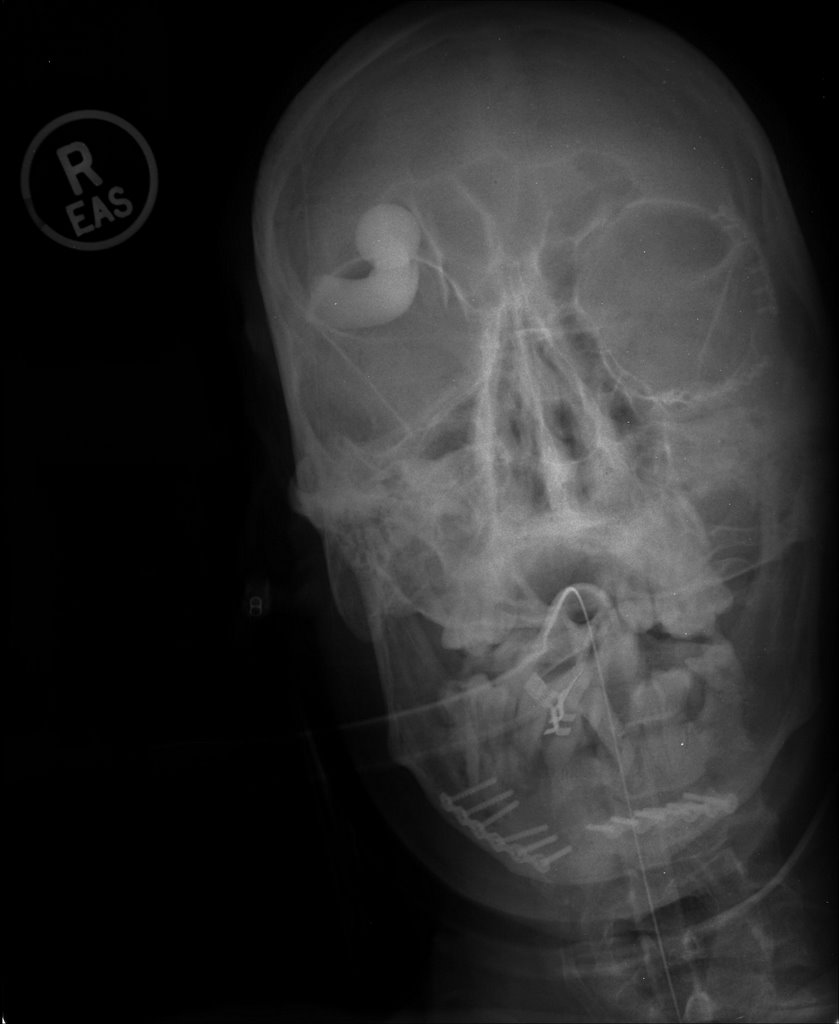
Something is here that doesn't belong. From the plates inidcated by the yellow lines
we can see that this patient has been a guest of the trauma service before. More images to follow:
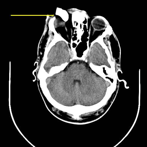
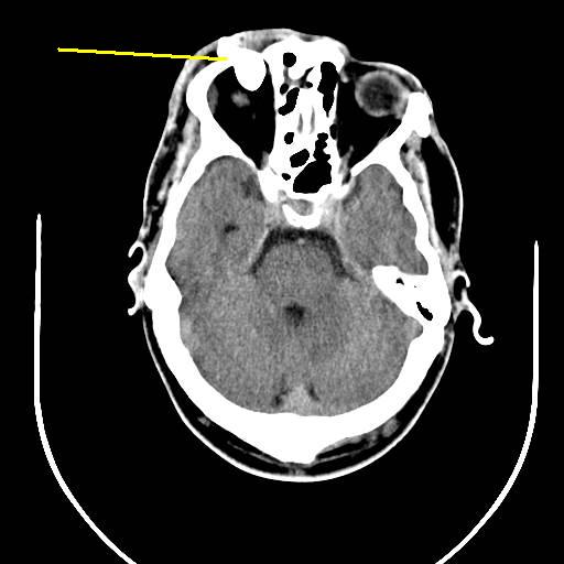


Brain windows on the head CT. Whatever it is it appears to pass over the globe and enter the frontal lobe with associated hemorrahge. Here is the bone window of the third image:

You can make out the fracture of the orbital roof. Here are the recontstructions from the facial CT:
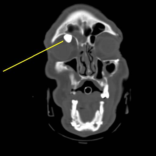

Here's the best of these:

And now for the really cool images:

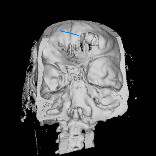
These are from the 3-D recons. The blue line indicates the offender. Here is the obligitory live action shot:
WARNING !!! THE FOLLOWING MAY BE OFFENSIVE:
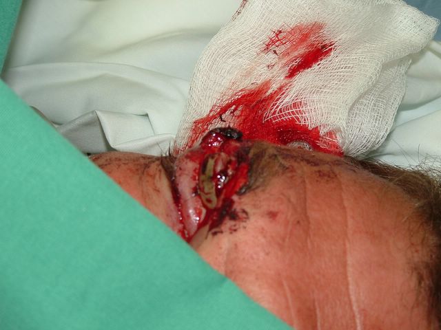
The object in question is just above the eyelid. The patient was taken off to the OR by the neurosurgeons for removal and placement of an ICP monitor. What was it? Probably the swivel mount from a rear-view mirror.
Labels: Tales from the Trauma Service
|Friday, October 13, 2006
Patient shot in the back at unknown range. Allegedly with a .223 caliber round. Here is the initial x-ray:

The yellow line (and paperclip) indicates the entry wound on the patient's back. Now as for the exit, that will be revealed shortly. Here are some additional images:

The yellow line represents a lamina fracture of the first thoracic verterbra. No hints on what the green line points to.
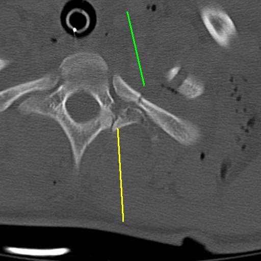
Transverse process fracture (yellow) and an associated rib fracture (green).
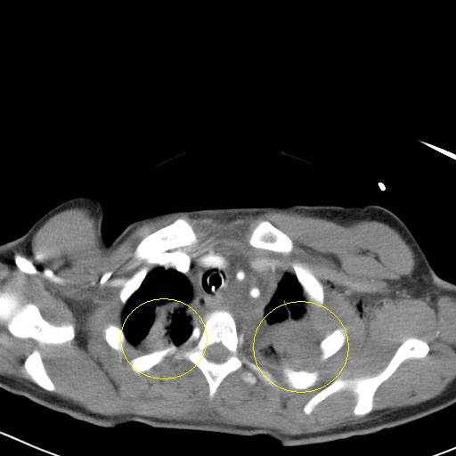
Bilateral pulmonary contusions, left greater than right. There is also some blood in the left hemithorax.
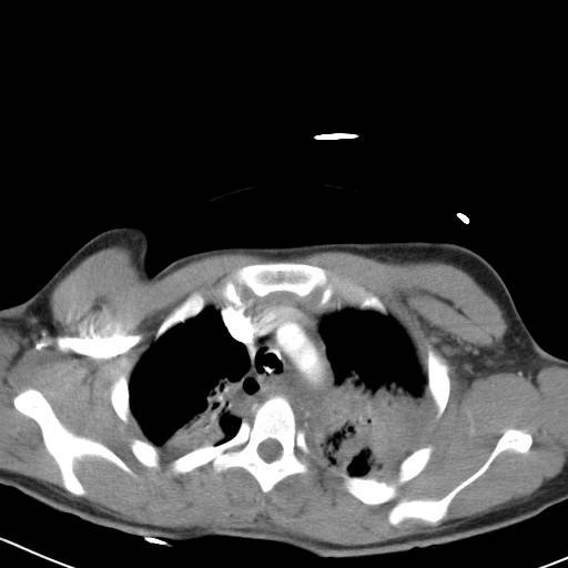
The contusions continue. Now for what you have been waiting for:
WARNING, POTENTIALLY OFFENSIVE IMAGES FOLLOW:
YOU HAVE BEEN WARNED!!!
Here is the exit on the anterior neck:
With a 6cc syringe for comparison:

The structure at the anterior of the wound is the posterior border of the sternocleidomastiod (SCM) muscle. I was able to digitally explore the wound and able to palpate inferior to the fractures seen in the CT scans above. Bleeding was primarily from fractured bone and torn muscle. No injury of the carotid, jugular vein, esophagus or trachea was seen. The wound was irrigated, packed with gelfoam and surgicel and I placed a Jackson-Pratt drain (that's what the green line was indicating).
But something very important does live there:
The brachial plexus. Here is some of the anatomy of the area:

From this diagram we can see the scalene muscles which lie posterior to the SCM. Those were pretty much destroyed by the blast. This next diagram

The brachial plexus also lies here. The phrenic nerve (which controls the diaphragm) does too. Prior to intubation the patient had no motor or sensory function of their left arm. We will probably obtain an EMG next week.
Labels: Tales from the Trauma Service
|Friday, August 11, 2006
This guy needs to buy himself a lottery ticket. The injuries you are about to see are from a fight over fifteen dollars worth of crack cocaine. If that is what the street value of life is these days.....
This guy also drove himself to the hospital.
Start the film:


Big big knife. That's gonna leave a mark.....
As he was stable my partner who was on call obtained a CT of his chest.
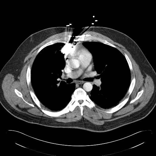
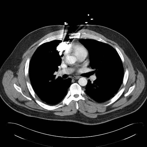
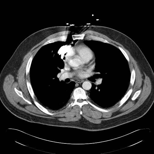


Minimal pneumothorax, minimal to no hemothorax, no fluid in the pericardium and the edge of the knife just millimeters from the pericardium. My partner and the local thoracic surgeon take the patient to the O.R. He is under general anesthesia, a chest tube placed and with cell saver, sternotomy saw, and 8 units of PRBC at the standby the knife is removed...........
Nothing happens. An occlusive dressing is placed on the wound. He ought to go home in a few days.
Labels: Tales from the Trauma Service
|Wednesday, February 01, 2006
Young man is a unrestrained back seat passenger in a MVC. CT shows an small intracranial bleed. No other injuries are found. He is awake and alert. A foley is not placed, and since this was one of my partner's cases, I am unsure if he underwent one of those new-age evaluations. He then began to complain of urgency and the inability to void. A foley was unable to be passed and his proximal thigh began to swell. Based on all this a cystogram was ordered:

The black lines represent the CT contrast in his bladder. The blue line represents extravasation of contrast from a urethral injury. He underwent bedside percutaneous cystostomy tube placement and was discharged to home with urologic follow-up on postinjury day six.
The mechanism? Probably the so-called "straddle injury" seen here:
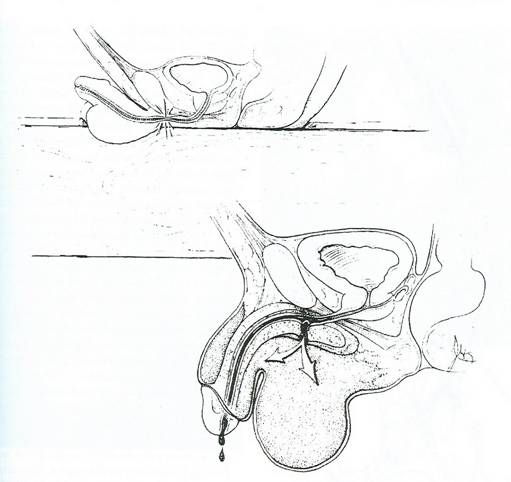
The urethra is trapped between the symphisis pubis and an inflexible object. This injury can result from playground injuries as well.
Labels: Tales from the Trauma Service
|Friday, January 06, 2006
Twenty-ish year old unrestrained driver in MVC. Airlifted from the scene. Intubated with difficulty in the ED. Difficult to oxygenate with a O2 saturation in the 40 to 50 percent range. Here is the CXR:
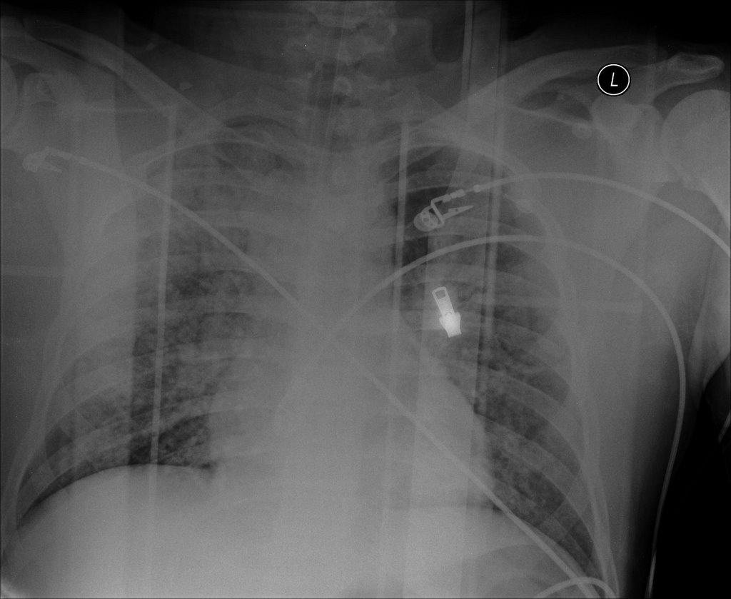
After some vent maneuvers such as increasing PEEP and using pressure control ventilation we are able to raise his saturation to the 70 percent range so we can go to CT:
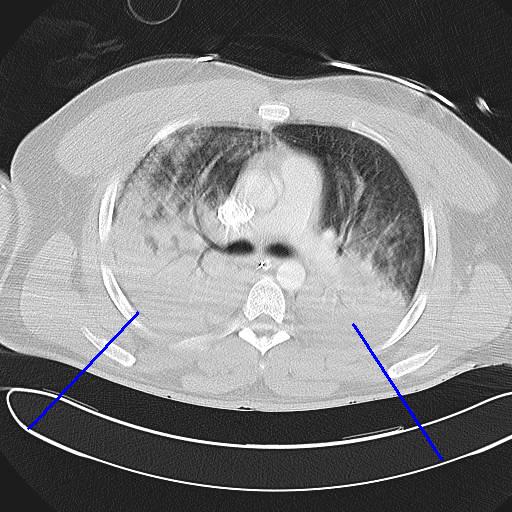
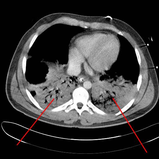
As you can see there were severe contusions bilaterally. No fluid in the posterior and there is no evidence of a pneumothorax. To add to this young man's woes:

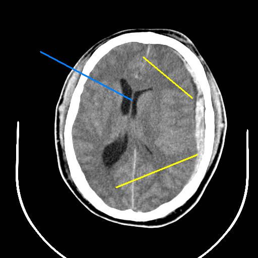
He has a severe subdural and subarachnoid hemorrhage (yellow) and a midline shift (blue. The enemies of head injuries are hypotension and hypoxia. After discussion with the neurosurgeon and anesthesiologist the decsion is made to take him to the OR for a craniotomy and chest tube placement. Chest tubes were placed because of the potential for barotrauma with high pressure ventilation. Try as we might, it took about 16 hours to correct his hypoxia. When oxygen is restored to a previously hypoxic organ (or blood flow is re-established) you can get what is known as a repefusion injury. As oxygen is restored there evolved into what could be described as a body wide re-prefusion injury with hypotension that required high levels of pressor support. His neurologic status continued to decline and his family decided to de-escalate his care and he expired on the fourth hospital day.
Again, somedays you get the bear, somedays the bear gets you.
Labels: Tales from the Trauma Service
|Monday, December 19, 2005
Every once in awhile you come across a radiographic finding that looks "just like in the book".
Young man falls from a second story window. Arrives somnolent but will awaken to voice. Multiple episodes of nausea and vomiting.
The images:
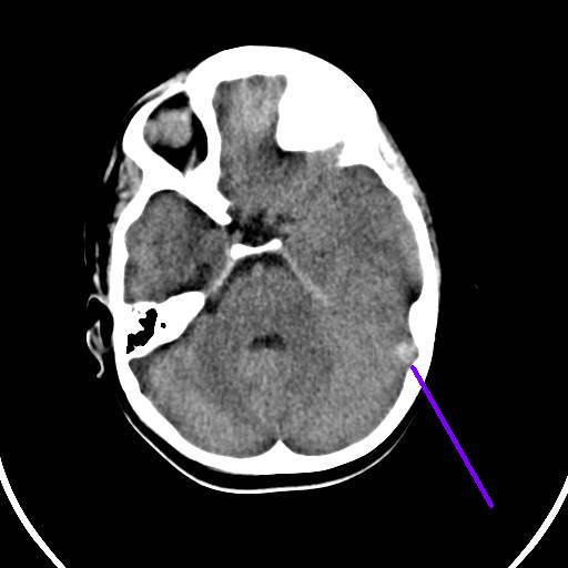
Small parenchymal bleed.

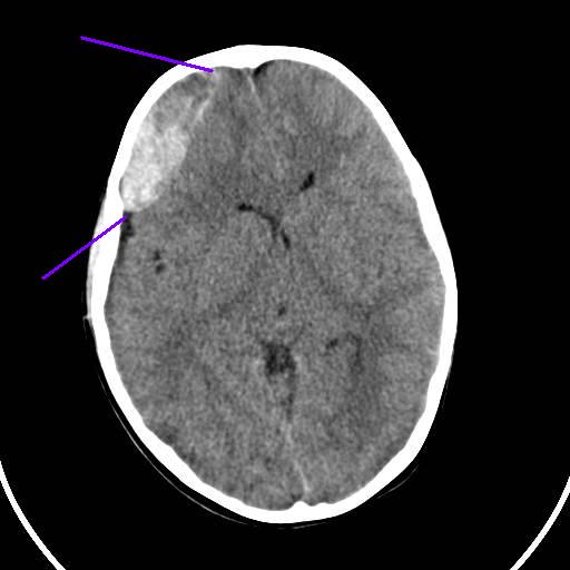

The classic radiographic appearance of an epidural hematoma. The neurosurgeon took him to the operating room for a decompressive craniotomy.
Labels: Tales from the Trauma Service
|Monday, October 17, 2005
A post a few weeks in the making. 20-ish man in a motor vehicle collision. Open right elbow fracture and a severe degloving imjury to his calf.
After some debridement here is what I was left with:

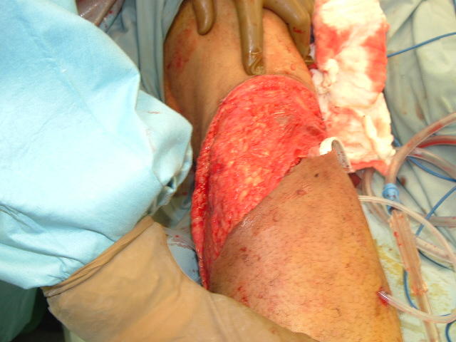
A bit more distal from the drain you see on the right was the extent of the degloving. I washed out the flap with about 3 liters of antibiotic containing saline. The drains were placed and the free edge sutured to the fasica with interrupted Vicryl sutures.
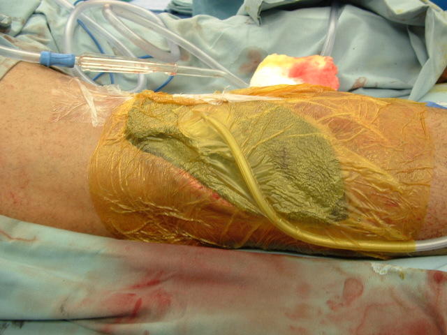
Over the open defect I placed a VAC dressing. The whole leg was then wrapped with an ACE bandage and he was placed in a knee immobilizer. After about 10 days of QOD trips to the OR for dressing changes under anesthesia it was finally time to graft. Here is what it looked like on that day:

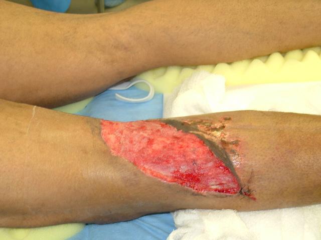
I debrided the inferior skin edge slightly and took the grafts from his poterior thigh. I also removed the JP drains. Applied another VAC over some Adaptic and let it sit for four days. Here is what it looked like:

Close to a 100% take. He is out of the knee immmobilizer now and ambulating.
Labels: Tales from the Trauma Service
|


