Thursday, November 11, 2004
Tales from the Trauma Service.....
I've been posting mostly on social and other issues, while holding back on what people really visit this site for....posts with lots of cool pictures!
This was one of my partner's cases, but interesting. A teenaged girl, pregnant BTW, shot by a known assailant in the left temple. Entry just posterior to the left orbit. Here is a cut from the facial CT:
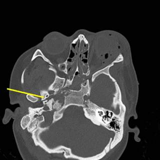
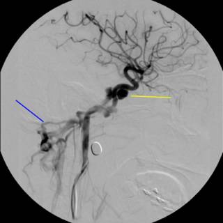
As you can see, the right wing of the sphenoid bone is fractured. Take a moment to contemplate what lives there. We sure did. Initial carotid angiogram:
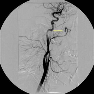
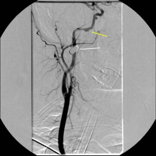
The white line indicates the bullet. The yellow line indicates what was thought to indiate a traumatic aneurysm or dissection. We sent the image to academic neuroradiologists and they agreed with the diagnosis. The debate then became what to do with it. Given the location of the vessel, operative therapy was ruled out. We were able to take a gradual approach due to the fact that that patient remained asymptomatic. Anticoagulation was considered, to prevent any thrombus formation and propagation, but the patient had a small epidural hematoma. Repeat angiography....
Oooohh! Much bigger now, and for the parting gift...
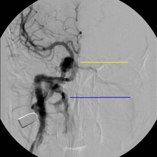
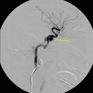
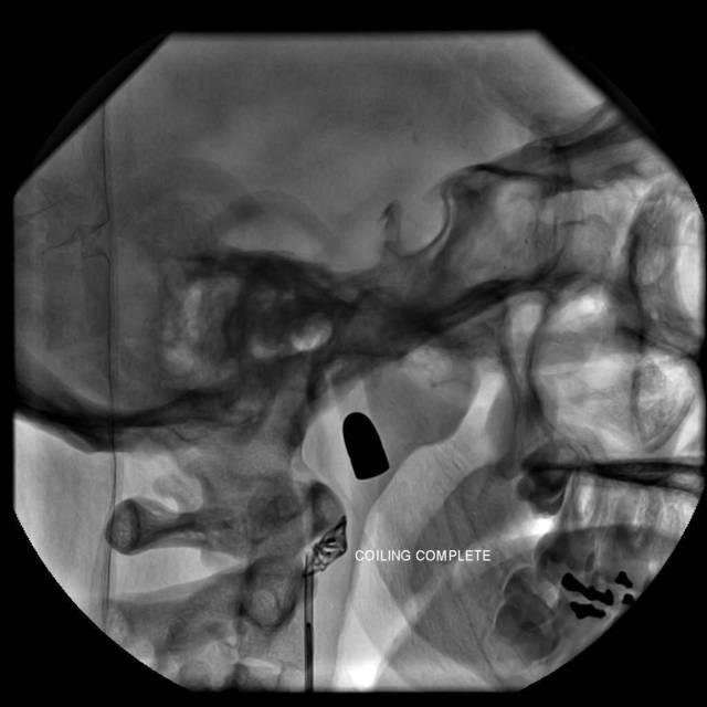
What we have is a traumatic arterio-venous fistula. So our hand was kind of forced. The option of coil embolization was entertained at the beginning, but sort of a "hot potato" that no one wanted to touch. After we determined the patient had an intact circle of Willis, a balloon was inflated in the proximal internal carotid artery, and held in place for fifteen minutes. After a few rounds of "Mary had a Little Lamb" and squeezing a ball without difficulty, the problem was addressed:

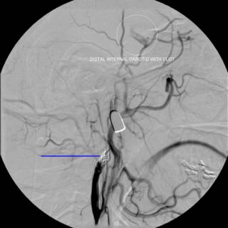
As you can see from the last angio there was alreadt thrombus present. This case shows the advantage of youth and an intact circle of Willis. She was discharged and is doing well. |
I've been posting mostly on social and other issues, while holding back on what people really visit this site for....posts with lots of cool pictures!
This was one of my partner's cases, but interesting. A teenaged girl, pregnant BTW, shot by a known assailant in the left temple. Entry just posterior to the left orbit. Here is a cut from the facial CT:

As you can see, the right wing of the sphenoid bone is fractured. Take a moment to contemplate what lives there. We sure did. Initial carotid angiogram:


The white line indicates the bullet. The yellow line indicates what was thought to indiate a traumatic aneurysm or dissection. We sent the image to academic neuroradiologists and they agreed with the diagnosis. The debate then became what to do with it. Given the location of the vessel, operative therapy was ruled out. We were able to take a gradual approach due to the fact that that patient remained asymptomatic. Anticoagulation was considered, to prevent any thrombus formation and propagation, but the patient had a small epidural hematoma. Repeat angiography....

Oooohh! Much bigger now, and for the parting gift...


What we have is a traumatic arterio-venous fistula. So our hand was kind of forced. The option of coil embolization was entertained at the beginning, but sort of a "hot potato" that no one wanted to touch. After we determined the patient had an intact circle of Willis, a balloon was inflated in the proximal internal carotid artery, and held in place for fifteen minutes. After a few rounds of "Mary had a Little Lamb" and squeezing a ball without difficulty, the problem was addressed:

As you can see from the last angio there was alreadt thrombus present. This case shows the advantage of youth and an intact circle of Willis. She was discharged and is doing well. |
![]()


