Tuesday, December 07, 2004
Tales From the Operating Room V.....
20-ish postpartum female with several bouts of biliary colic during pregnancy. Set up for cholecystectomy. Liver function tests normal. No jaundice or any other signs of biliary obstruction. Intraoperative picture:
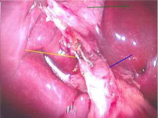
The green line points to the gallbladder, the yellow line to the cystic duct with clips on it and the blue line to the common bile duct, which looked enlarged in my opinion. This picture was taken after the cholangiogram was performed. I perform my cholangiograms with a cystic ductotomy and an Arrow cholangiocatheter. Here are the images:
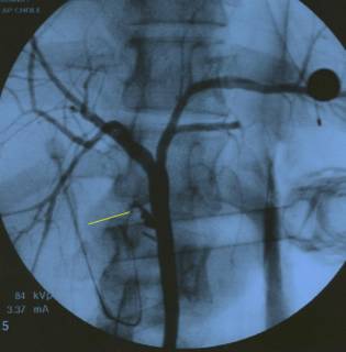
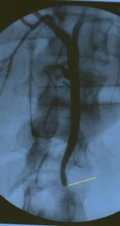
The proximal system fills well. Yellow line points to the cystic duct. The common duct does not appear to be too dilated.
Here's the rub. Normally you would see contrast flowing to the duodenum, indicating the duct was clear. As seen in this study from another patient:
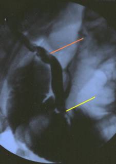
The yellow line shows contrast within the small bowel. The red line shows stones within the common duct.
The differential in this case is: 1) retained common duct stone, 2)malignancy, which would be uncommon in an otherwise healthy 20 year old, and 3)Sphincter of Oddi spasm. The radiographs did not change after administration of glucagon and 20 minutes of breathless anticipation.
This is one of those times that the old "surgical judgment" comes in to play. What to do? Open common bile duct exploration? Laparoscopic ? Both of those are limited by the fact the duct is not that large. Such a duct would be difficult to manipulate and could stricture, causing later trouble. Fortunately I have some excellent gastroenterologists in River City and the patient underwent a successful endoscopic retrograde cholangiopancreatography (ERCP) the next day with a sphincteroplasty. (Sorry, the ERCP pictures aren't very good). Several stones were extracted and she was discharged. |
20-ish postpartum female with several bouts of biliary colic during pregnancy. Set up for cholecystectomy. Liver function tests normal. No jaundice or any other signs of biliary obstruction. Intraoperative picture:

The green line points to the gallbladder, the yellow line to the cystic duct with clips on it and the blue line to the common bile duct, which looked enlarged in my opinion. This picture was taken after the cholangiogram was performed. I perform my cholangiograms with a cystic ductotomy and an Arrow cholangiocatheter. Here are the images:
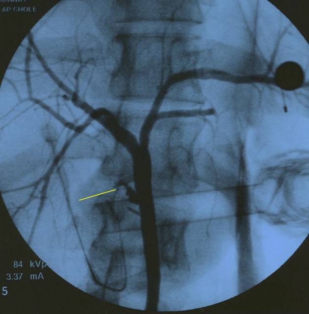
The proximal system fills well. Yellow line points to the cystic duct. The common duct does not appear to be too dilated.

Here's the rub. Normally you would see contrast flowing to the duodenum, indicating the duct was clear. As seen in this study from another patient:

The yellow line shows contrast within the small bowel. The red line shows stones within the common duct.
The differential in this case is: 1) retained common duct stone, 2)malignancy, which would be uncommon in an otherwise healthy 20 year old, and 3)Sphincter of Oddi spasm. The radiographs did not change after administration of glucagon and 20 minutes of breathless anticipation.
This is one of those times that the old "surgical judgment" comes in to play. What to do? Open common bile duct exploration? Laparoscopic ? Both of those are limited by the fact the duct is not that large. Such a duct would be difficult to manipulate and could stricture, causing later trouble. Fortunately I have some excellent gastroenterologists in River City and the patient underwent a successful endoscopic retrograde cholangiopancreatography (ERCP) the next day with a sphincteroplasty. (Sorry, the ERCP pictures aren't very good). Several stones were extracted and she was discharged. |
![]()


