Wednesday, June 29, 2005
Do You Know Where that Finger has Been??????
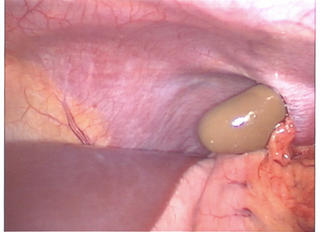
A follow-up to this post. The one that had this picture:

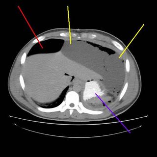
The patient progressed slowly with low-grade fevers and leukocytosis. He had persistent free air as well so after a few days I obtained a CT Scan:


The yellow lines are a subphrenic fluid collection, likely an abscess. The red line is more free air and the blue line is contrast in the stomach. Interventional radiology again saves the day with a pigtail catheter.

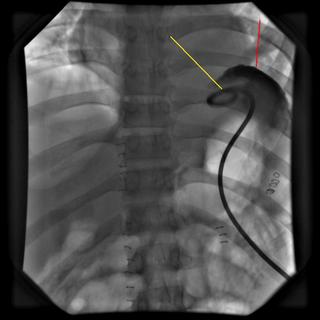
The yellow line points to the catheter, red demonstrates contrast within the cavity. About 1500cc are removed initially with decrease in WBC and the patient becomes afebrile. A repeat scan is obtained a few days later:
The patient had received 24 hours of Ancef postop. The cultures grew out S.viridans so a missed visceral injury is unlikely. He was taking a regular diet throughout the above events. He was discharged a day after the repeat CT. I saw him recently in the office and he is doing well. |
A follow-up to this post. The one that had this picture:

The patient progressed slowly with low-grade fevers and leukocytosis. He had persistent free air as well so after a few days I obtained a CT Scan:


The yellow lines are a subphrenic fluid collection, likely an abscess. The red line is more free air and the blue line is contrast in the stomach. Interventional radiology again saves the day with a pigtail catheter.

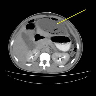
The yellow line points to the catheter, red demonstrates contrast within the cavity. About 1500cc are removed initially with decrease in WBC and the patient becomes afebrile. A repeat scan is obtained a few days later:

The patient had received 24 hours of Ancef postop. The cultures grew out S.viridans so a missed visceral injury is unlikely. He was taking a regular diet throughout the above events. He was discharged a day after the repeat CT. I saw him recently in the office and he is doing well. |


