Tuesday, August 24, 2004
Scan' em all and let God sort them out...
Had planned a lengthy rant today on an issue of import, but silly day job reared its' ugly head. Anyway, a brief discussion of how the use of thoracic CT is expanding.
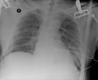
70-ish restrained passenger struck on their side of vehicle. +LOC. GCS 15 0n arrival. Main complaint is of back pain. Physical exam unrevealing. Initial CXR:

Not much to see. But as my use of FAST has reduced the number of abdominal CTs I order on trauma patients, the number of chest CT's I order has increased. From the May 2002 Journal of Trauma: Incidence and Characteristics of Motor Vehicle Collision-Related Blunt Thoracic Aortic Injury According to Age.
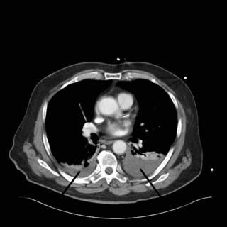
I did perform a FAST exam, and I could not get good visualization of the splenorenal space very well. Off to CT. Solid organs look O.K., but look what we found:

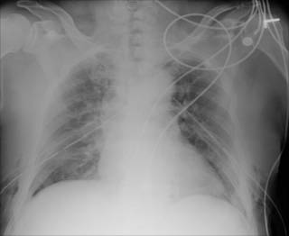
There is about 500cc of blood on the left and 75cc on the right. Here is the dual exhaust film:

While the hemothorax was not hemodynamicaly significant, blood left in the chest can lead to empyema, fibrothorax, and entrapment of a lobe. These conditions can lead to a thoracotomy and decortication. Not fun.
See this post from Shazam for more CT scanning adventures.
|
Had planned a lengthy rant today on an issue of import, but silly day job reared its' ugly head. Anyway, a brief discussion of how the use of thoracic CT is expanding.
70-ish restrained passenger struck on their side of vehicle. +LOC. GCS 15 0n arrival. Main complaint is of back pain. Physical exam unrevealing. Initial CXR:

Not much to see. But as my use of FAST has reduced the number of abdominal CTs I order on trauma patients, the number of chest CT's I order has increased. From the May 2002 Journal of Trauma: Incidence and Characteristics of Motor Vehicle Collision-Related Blunt Thoracic Aortic Injury According to Age.
Abstract:But submitting Grandma and Grandpa to thoracic angiograms is not a benign course of action. The use of thoracic CT has made the diagnosis of the injuries simpler.
Background : Motor vehicle collision-related blunt thoracic aorta injury (BAI) is rare and highly lethal. Vascular disease as related to advancing age potentially subjects older adults to increased risk of BAI; the mechanisms associated with such injuries may be different as compared with younger adults. The goal of the present study is to test this hypothesis using population-based data.
Methods : The 1995 to 1999 National Automotive Sampling System data files were used. The National Automotive Sampling System is a national probability sample of passenger vehicles involved in police-reported tow-away crashes. BAI was defined according to the Abbreviated Injury Scale codes. Among those with BAI, information on occupant (age, seating position, restraint use), collision (collision type, delta-V, vehicle intrusion), and outcome characteristics were obtained and compared according to age.
Results : The overall incidence of BAI was 6.8 per 10,000 occupants and there was a steady increase in the BAI rate for advancing decades of life. The proportion of occupants with BAI who die at the scene of the collision is relatively consistent across all age groups (~85%). Among those who survive to receive medical care, ultimate survival is lowest among those aged 60 and older. Near-side collisions were responsible for more BAI among older adults than other age groups (50% vs. 20.6%, p <= 0.05). Older adults sustained BAI in collisions with lower delta-V values compared with younger persons (p <= 0.05). Conclusion : Older adults have the highest rate of motor vehicle collision-related BAI, and their injuries tend to occur in less severe collisions. A high level of suspicion for BAI among older adults should not be reserved for high-energy collisions only.
I did perform a FAST exam, and I could not get good visualization of the splenorenal space very well. Off to CT. Solid organs look O.K., but look what we found:

There is about 500cc of blood on the left and 75cc on the right. Here is the dual exhaust film:

While the hemothorax was not hemodynamicaly significant, blood left in the chest can lead to empyema, fibrothorax, and entrapment of a lobe. These conditions can lead to a thoracotomy and decortication. Not fun.
See this post from Shazam for more CT scanning adventures.
|


